


 시약 및 소모품
 이미지분석장비
Omega Lum™ C
Omega Lum™ C Imaging System
Quantitative Western Imaging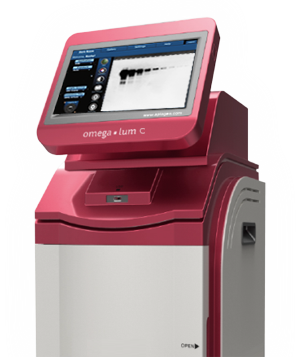
Contact us today and request an instrument for your free one week trial.
Quantitative Results
The Omega Lum C is optimized for superior detection of chemiluminescent Western blots. A wide dynamic range and fast imaging speeds mean that you will spend less time trying to get the perfect image.
Dynamic Range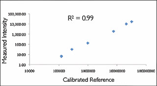
Sample: Calibrated luminescent plate
Imaging Method: Chemi-HiRes
Dynamic Range: 3.4 orders of magnitude
High Resolution
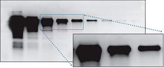
Sample: Purified transferrin
Membrane: PVDF
Primary antibody: Rabbit-anti-transferrin
Secondary antibody: Goat-anti-rabbit
Substrate: Advansta WesternBright Quantum
Imaging method: Chemi-Hi Res
Sensitive Detection
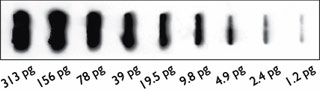
Sample: Slot blot of secondary antibody
Membrane: Nitrocellulose
Antibody: Anti-rabbit-HRP
Imaging method: Chemi
Imaging time: 4 minutes
Application Flexibility
multiple excitation sources allow you to visualize a wide range of gel stains, making the Omega Lum c a versatile imaging system.
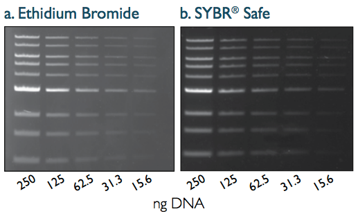
Sample: Quick-Load® 1 kb dnA Ladder (neb)
Gel: 1% agarose in TAE
Stain: a) Ethidium bromide, b) SYBR safe
Imaging Application: a) EtBr, b)SYBR safe
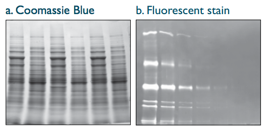
Protein gels stained with a) coomassie blue and b) a fluorescent protein stain. The Omega Lum c captures high resolution images for documentation or publications.
Sample: a) Whole cell lysate, b) protein purification
Stain: a) coomassie blue, b) Advanstain scarlet™
Imaging Application: a) coomassie blue, b) custom
Specifications
Camera : • 8.4 MP for high resolution images
• 16 bit scientific grade CCD camera for quantitative imaging
• Peltier cooling, -50ºC controlled for chemiluminescence imaging
• USB connection for fast downloads
• Fast refresh rate for live imaging
• High quality images ideal for downstream analysis
Lens : F 0.95 Lens
Field of View : 15 x 20 cm
cabinet : • 302 & 365 nm pull out UV transilluminator
• EPI white LEDs
• EPI blue LEDs
Filter Wheel : 6 Positions, motorized
Filters : Orange Filter (590 nm)
Applications : Chemiluminescent Western blots, fluorescent and visible gels, and SYBR® safe dye imaging
Certifications : CE, cTUVus
Product Footprint : 12.5” x 18” x 27.5” (31.75 x 45.72 x 69.85 cm)
